The ESC Digital congress was a bold and
successful initiative of the European Society of Cardiology, and took place in
Tallin, a few days ago. The location was not chosen by hazard, as Estonia has
99% of health data digitized and 99% of prescriptions are digital. Prof.
Viigimaa exposed the architecture of the Estonian Electronic helth system in a
comprehensive presentation
at the beginning of the congress.
Current organization
of digital healthcare
was reviewed by Prof. Martin Cowie in an excellent presentation,
where he highlighted that we need to identify barriers and find appropriate
solutions on this emerging sector. We will always find at the center of our
care the patient and his family, and this aspect should not be overlooked or
forgotten, especially in this rapid-digitalized era.
Patient care, patient empowerment
and nursing issues
were also addressed by Prof. Donna
Fitzimons and ACNAP President-Elect
Lis Neubeck, with accent on developing the short and longer path to
action which have in center the patient and the nurse (as midlevel team
member). Digital nursing sessions drawed a lot of constructive comments, with Donna Fitzimons opinioned that
if the patient is art ease at using online interaction with a “digital” nurse
(avatar talking and guiding the patient by means of AI), then we should use
this technology mainly to prevent, and not to treat. Find below the interesting
slides from the presentations and their link
to full presentations.
A very interesting session addressed the issue
of preparing the healthcare force for
the digital future chaired by ACCA President
Susanna Price. Even though some solutions have been highlighted in a
important scientific
paper, like as the need for a culture of learning (develop extensive
learning environment, encourage innovation and dare to fail), building a strong
learning infrastructure and developing a multi-professional and collaborative
approach to learning, these approaches are unachievably in the current state of
the medical system in Europe, where most of the current medical personnel are
disengaged or over-fatigued, doctors and nurses are burned-out.
Does technology make things worse for the
medical workers? Can artificial intelligence and automation ease things? See
all the debate here.
The same issue was addressed in another session.
While trying to find solutions for the future, present issues of healthcare
professionals and the digital boom relate to lack of trust and poor experience
with new technologies, difficulty to interact with technology, fear of change. This
is maybe where artificial intelligence could make the difference, as it can
improve healthcare efficiency and delivery and could replace human involvement
in some tasks of the medical industry, as suggested Dr. Casado Arroyo. However,
for the moment, present algorithms are not completely in their mature stage.
Cybersecurity was a hot topic. Dr. Avi Fischer
resumed some of the important cautions to take in reducing cybersecurity risks,
such as:
sharing responsibility between
stakeholders-healthcare facilities, patients, providers, and manufacturers of
medical devise;
cybersecurity should be a priority
during the design and development phase of the medical device and the issue
should be addressed in a “Bolt-On to Integrated” fashion;
if a vulnerability is found,
communication and coordinated actions between stakeholders and healthcare
facilities are vital. Development and involvement of government structures to
assure security is very important.
The participants also expressed the
urgent need for developing secured protocols to transmit data.
A special session to electronic medical record (EMR) can be accessed here.
The consensus was that even though EMR are an efficient data base that can help
health professionals in decision making, it is also time consuming. A new concept
that consists on “less keyboard, more patient contact” should be adopted,
because according to one study, for every hour
physicians provide direct clinical face time to patients, nearly 2 additional
hours is spent on EHR and desk work within the clinic day, and outside
office hours, physicians spend another one
to two hours of
personal time each night
doing additional computer
and other clerical
work. Dr. Nico
Bruining
highlighted an important limitation of EMR used in Europe (vs. US), notably in
Europe we use more than 30 languages that makes data collection difficult, and
that should probably be aimed by a future Horizon 2020 project.
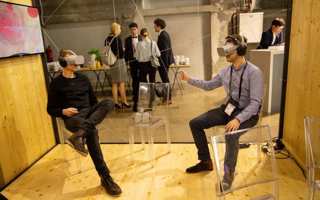
Augmented, mixed and virtual reality discussions were
divided into cardiologist-related and patient-related. The 3 terms are
intercorrelated and should not be confused one with another, here is their
meaning and a schematic representation:
Virtual reality (VR) immerses users in a fully artificial digital
environment.
Augmented reality (AR) overlays virtual objects on the real-world
environment.
Mixed reality (MR) not just overlays but anchors virtual objects to
the real world.
The 2 main applications
in medicine targets the patients and of course the doctors.
For patients, one
of the presenters suggested that VR will do for patient education what
Google Maps has done for navigation, thus it “will transform learning
experiences by better retention and recall, it will improve patient journey and
will determine behavioral change by challenging health beliefs through
impactful experiences”.
For doctors, Dr. Dariusz Dudek
explained how a pioneer programe of mixed reality that is currently used in
Poland helps cardiologists choose a better punction site for TAVI, with
expectancy in the very near future to have VR mask that will allow
cardiologists to have echo, CT and eventually other 3D reconstructions in the
corner of their eyes, while a cardiac intervention is taking place. Other
important applications are stroke recovery and cardiac rehabilitation.
Over-utilization of
medical devices.
One of the worries highlighted was that this
new technology could cause anxiety in healthy fit subjects, translating into
unnecessary consultations either by their general practitioner either by their
cardiologist, as presented by Prof.
Martin Cowie. 57% of subjects from the “Apple heart” study thought
to seek medical attention after having an alarm from a smart device.
Even though wearables and connected devices
impact sometimes positively our daily lives (sleep apnea analysis app, selfie
to quantify oedema app, accelerometers incorporated into devices that quantify
daily effort, smartphone-based blood pressure measurement by transdermal
optimal imaging, etc), Dr.
Klaus Witte highlighted that there is increased concern that
wearables and connected devices impact negatively the society by increased (but
unnecessary) cost to the consumer and increases (but unnecessary) cost to the
medical system (by over-investigating a healthy person thus launching a battery
of tests like holter ECG, echocardiography, stress test). With this in mind,
there was a unanimous consensus that medical wearables and connected devices
should target special groups of populations if we want to avoid death, strokes
or hospitalizations and the clinical benefit of these kind of devices should be
carefully thought and evaluated.
Conclusions
Digital technology can help healthcare by
providing new tools for diagnosis and therapy delivery, redesigning clinical
pathways, individualized risk stratification and individualized care, patient
empowerment, support to precision medicine (thus fewer medical errors), and
telemonitoring. Digital technology and connected devices (tablets, wearables,
apps) will enhance decision making of health professionals only if there is a
direct link towards a clinical benefit, and maybe they would have a huge impact
if they could be directly linked also to EMR. In real life, adoption of digital
technology in healthcare depends on validation, easy integration of workflow
& prescription pathways and ability to reduce work.
Consumers could think that by using digital
technology and connected devices, they take control over their health; this is
partially false because alarms issued by devices could translate into
unnecessary consultations and increased cost to overall health system by doing
unnecessary tests in otherwise healthy subjects. Narrowing the indications and
the target population for the use of this devices should be a priority.
And finally, some punctual issues need rapid
and clear solutions: stricter security protocols should be incorporated in data
transmission, regulations should be adopted for all digital technology (there
are too many devices and few regulations), the huge amount of data (that is
collected from all digital technology) needs to be transformed into
information.
According to an English study, 16% of the victims had been hospitalized in the previous month for atypical symptoms.
In England, one in six fatal myocardial infarction patients were hospitalized in the month before the diagnosis of infarction was reported by doctors. Could the fatal accident have been avoided? This is the question raised by the study conducted by Professor Perviz Asaria and his colleagues at Imperial College in London, together with researchers from Harvard, USA. The results were published in The Lancet Public Health. The researchers studied the medical record of the 135950 people aged 35 and over who died of a myocardial infarction in England between 2006 and 2010. They found that half had already been hospitalized in the four weeks before the accident Fatal, or a group of 66490 patients.
Missed opportunities or classification errors
They then dismissed two-thirds of those diagnosed with myocardial infarction, either directly or secondarily, and concentrated on the 21677 patients who were not thought of. For the most part (59%), the reasons for hospitalization did not particularly favor a cardiac problem (pneumonia, cancer, infection, fracture of the femur neck, etc.). But for 7566 patients (35%), the diagnosis made during the hospitalization was that of another cardiac disorder, including heart failure or rhythm disorder (atrial fibrillation in particular). In 1368 patients (6%), there were also symptoms suggestive of myocardial infarction, such as shortness of breath or atypical chest pain.
The fact that symptoms are underestimated does not mean that it is a automatically a medical error, but the study shows that the medical profession needs to improve risk stratification to better detect who is at risk of dying from an infarction in the event of atypical symptoms.
Establishing a prognosis is always more difficult than making a diagnosis a posteriori, and there is no question of keeping in the hospital all the people complaining of a gastric embarrassment
As for the chest pain typical of the infarction, a study conducted in North Carolina between 1994 and 2006 showed that it was absent in two thirds of infarcts. Doctors are very good at treating heart attacks when they are the primary cause of admission to the hospital, but much less when they are comorbid (pathology associated with another diagnosis, or when it comes to identifying subtle symptoms that could lead to the imminence of a myocardial infarction.
Failure in detection
These results should encourage physicians to be more vigilant, reduce the risk of missing these symptoms and ultimately save lives.
In France, it is estimated that 120000 infarcts (or acute coronary syndrome) occur each year. Improvements in the management of patients with chest pain have significantly reduced myocardial infarction mortality. But margins of progress still exist, as one-third of patients lose time by visiting their family doctor or by going to their own emergencies instead of directly calling the 112 or 15 emergency service, when they experience typical symptoms infarction (ongoing chest pain).
The chances of long-term survival increase significantly with prompt intervention following a cardiac arrest.
Every year in France, 40,000 people suffer from cardiac arrest. In Paris the chances of survival at 30 days were close to 10% in 2016, compared with 6% five years earlier. In Denmark, a country with 5.6 million inhabitants, the national registry’s rate of survival was 12% in 2013, compared to 5% in 2001. In the meantime, training of general population in resuscitation gestures and protocols in order to restart the heart was performed (cardiac massage started by a witness immediately after calling a specialized team, defibrillation if necessary). The result was a rise of the efficacy of resuscitation maneuvers from 18% in 2001 to 60% in 2013, according to the study published in the European Heart Journal. A key factor in increasing the survival of victims, survival which increased by 10%. It is important to note that the increase in resuscitation gestures by controls is not the only reason for this improvement. Quality care provided by emergency services and hospital care add to these results.
Since 2013, there has been a consensus within the Danish Cardiovascular Society that all victims of cardiac arrest whose heart is restarted (“recovered cardiac arrest”, in medical jargon) should benefit rapidly from a coronary angiography (examination to visualize the heart’s arteries), and, if one of them is occluded, a gesture to restore blood flow (angioplasty with stent). This means that they are brought by the emergency services directly to a hospital with a technical interventional cardiology platform.
By taking the 41,000 cardiac arrests collected in the Danish register between 2001 and 2013, Dr. Tranberg and his colleagues observed that 16000 patients had died before arriving to a hospital, 18000 were sent to local hospitals and 7000 were taken to an interventional cardiology center. The rate of survival was 29%. However, the study shows that the remoteness of the center does not significantly influence the survival rate. Once cardiac arrest is recovered, you can afford to lose a little time to go to a particularly experienced cardiac center.
In France, the transfer to a center with an interventional cardiology unit has already been carried out almost systematically for several years, but the quality of the hospital’s resuscitation service must also be taken into account, because the in-hospital management is almost as important as the initial phase of the recovery.
Regardless of the quality of medical services of the hospital where a cardiac arrest victim are transferred, the first few minutes are crucial. All adults should have the basic notions how to perform cardiac massage. Even before the transfer to a hospital, the most important thing is the initial management: the duration of the cardiac arrest before the first massage and the time taken to recover an effective cardiac rhythm.
High blood pressure, how to treat it, what hypertension means and how it affects us are questions that can be found in varied variants of answers, but the medical correctness of these types of questions can be found in this post.
Blood pressure represents physics laws as any fluid inside a container and exerts pressure on its walls. The fluid we are talking about is the blood that is in permanent circulation in the blood vessels and is constantly exerting tension on them.
The tension we are talking about is blood pressure (BP), which is the pressure exerted inside the blood vessels. Blood circulation has two values due to myocardial contraction and relaxation, therefore blood pressure is expressed in two sets of values: one higher (systolic) and a smaller one (diastolic).
Blood pressure is measured in mm / hg, meaning millimeter mercury column, and the abbreviated form is to divide the values at 10, so the correct expression is like an example of 120/80 mm / Hg while the abbreviated form is 12/8.
Normal range.
Blood pressure values can not be categorized into a clear figure, but rather within an interval because blood pressure varies as normal limits depending on:
– Weight: A person under 55 kg has a value of 10 mm / Hg at lower acceptable limits, while a person with a value between 55-85 is exactly the average of the values, and between 85-110 is accepted by 10 Mm / Hg above the admitted limit, as in people over 110 kg most likely will be a obesity hypertension, that is to say even at a value of 20-30 mm / Hg but which is not necessarily accepted; some expect increased value in these people with obesity;
– Age: For young adults aged 16-25, the tension may be either exactly the normal range or exactly 10 units below this average. For the mature adult: 25-45 years of age should fit exactly within the medium range. While between 45 and 60 years, this value is even 10 units over this range, and over 60 years there may be hypertension or hypotension (so-called physiological), that is, 10, even 20 units above the mean .
– Physical Conformity: If a physical person is athletic conformation or practicing regular sports, or has practiced a performance sport for more than 10 years and has not passed more than 10 years since the end of performance sports, a Adaptability of the heart (as a rhythm) a decrease in this rhythm and respiratory rate as well as blood pressure, so it is possible for these people to have a value of less than 10 in the category of the person.
– In these characteristics where small varieties can be accepted, there can be added also environmental and environmental conditions: smoking, alcohol consumption. Psychosocial factors can also be added: stress, psychic shock recently. At the same time, a physical exercise may have to be considered that may have been given up before the voltage measurement (less than 5 minutes before blood pressure measurement).
A correct measurement to confirm the certainty of possible blood pressure pathologies should include three consecutive measurements at a distance of 3-5 minutes apart, alternating the two arms.
Hypotension and Hypertension – Values, Explanations, Symptoms
For clarification, an average value for a young adult or mature adult is 90-139 mm / Hg of systolic and 60-89 of diastolic pressure.
Hypotension is the condition of blood pressure below normal values, i.e. below a systolic value of 90 and an adjacent one, diastolic below 60 mm / Hg. If this tension is in the range of -10 mmHg to both systolic and diastolic, and the person concerned is either a female patient with a body weight of 40-45 kg (or in some cases even more If the person is athletic performance or if the person’s fluid consumption is very low, there are already possible explanations for the hypotension.
A liquid consumption of less than 1 liter per day (although physiological consumption is 2-2.5 liters of fluid per day than those in food or other forms of liquids rather than water), is sufficient to favorize hypotension. Blood pressure is directly proportional to the circulating blood volume (a smaller amount of blood will exert a lower pressure on the walls).
Symptoms related to hypotension are: fainting, dizziness, feeling of choking that can be associated with a headache, dyspepsia (indigestion) or dysuria (painfur urination), and can progress to the loss of consciousness .
High blood pressure first class means an increase over the value of 140/90 of blood pressure and that can explain a heart failure at the onset of pathology, especially if the systolic value is 140 and the diastolic is 95-100. But in time, it has two paths, or it will be adapted by the body that will be able to compensate it, ie to adapt to these values, or it will evolve to grade 2 hypertension. A grade one hypertension will give in extremely rare cases complications that are generally referred to as hypertension, but progressive progression through second degree will later give these complications. Symptoms of the first degree hypertension include palpitations (which may also indicate associated cardiac dysfunction), a headache, or vertigo and pressure in the ears or in the eyelid.
Second-degree hypertension is already an important hypertension that needs to be treated and that already reveals some dysfunctions: cardiac, renal, or circulatory. This hypertension can cause complications such as: aneurysm (aortic or abdominal artery aneurysm), heart failure, chronic kidney pathologies, cardiac arrest or infarction as well as peripheral arterial pathology as well as coronary artery disease. Symptoms include tinnitus (headache), headaches (especially frontal), vertigo and dizziness as well as episodes of fainting.
Third-degree hypertension is a medical emergency, meaning a major dysfunction. Acute complications include stroke, heart attack, heart failure. hronic complications include: heart failure, coronary artery disease or thoracic/abdominal artery aneurysm.
Causes
Causes of hypertension originate from a combination of interactions between several factors, most of which are factors that have been acquired (living and environmental conditions also):
– Smoking and alcohol consumption: Although it acts by two different mechanisms, smoking causes both a respiratory and endothelial dysfunction that will also cause hypoxia and a deficient oxygen fixation on hemoglobin. However, alcohol will act through portal hypertension (portal system of the liver) and the portal hypertension will cause hypertension.
– Obesity: Obesity will form plaque atherosclerosis in the blood vessels and which in turn will aggravate blood circulation through the vessels. This will result in high blood pressure.
– Stress: In addition to a series of strict cardiac and cardiovascular pathologies other than hypertension, and an aggravating cause in ulcerative pathology, stress is a major incriminating factor in hypertension as well.
The main two organs that can favorize hypertension are the heart and the kidneys.
Pathologic vessels, i.e. weakening of vessel elasticity caused by deposition of atheromatous plaques, a decrease in elasticity due to age, or even clot formation can cause hypertension.
If the catheter was inserted in the arm, at the end of the procedure the catheter and sheath are removed. The incision will be closed through compression bandages. You will be forced to keep the arm still for at least one hour. You will be observed for several hours to ensure that you feel good after the procedure. You may receive medication to relieve discomfort in the arm after the anesthetic effect disappears. You will be given instructions on how to take care of your arm when you return home. Tell your nurse if you see blood or feel any numbness or tingling sensations in the fingers.
If the catheter was inserted in the groin at the end of the procedure catheter and sheath are removed and the incision is closed by compression or with a “plug” of collagen (accelerates the formation of a clot in the artery), and then bandaged . A sterile dressing and compression will be put on the groin area to prevent infection or bleeding. You will need to not move position of the foot for 3-6 hours in order to prevent bleeding. Head can not be raised more than the level of pillows (about 20 degrees). Do not raise your head off the pillow (try not to look ahead), as this can contract the abdominal muscles and can promote bleeding from the puncture site. Do not try to stand. Nurse will check the bandage regularly. Warn her if you see blood orif you experience a sensation of humid, warmth at the punction site, or if your toes begin to tingle or feel numb. You may receive medication to relieve discomfort in the groin after anesthesia passes.
Your nurse will help you step out of bed when you are allowed to get up (after at least 6 hours of stretched leg position). Ideally, you should stay in bed 12 hours. You will then be allowed to get out of bed, you can go to the bathroom. If you need assistance seek the help of a nurse. An video of the exam can be found here.
You will need to drink plenty of fluids to remove the dye in the body. You may feel the urge to urinate more frequently. This is normal. If a urinary catheter was not placed during the procedure, you have to use a urinal until you are able to get up from bed.
Your cardiologist will tell you if you are able to return home or will have to stay overnight. Usually after a “radial” puncture is not required staying overnight in the hospital unless you experience chest pain or heart arythmias. Treatment, including medications, dietary changes, and future procedures will be discussed with the doctor before going home. You will also talk to your doctor, says Alexandru Mischie about proper wound care, your work, but also about recovery and whaterver subjects you have in mind.
A nurse will insert an intravenous line in your arm so that medications and fluids can be administered through the vein during the procedure. The nurse will clean the skin (and possibly shave), the place where the catheter will be inserted (arm or groin). Sterile fields are used to cover the site and help prevent infection. It is important to keep your arms and hands to your sides and not touch the sterile field. Electrodes (small, flat, sticky patches) will be placed on the chest. The electrodes are attached to an ECG device that provides electrical diagram of the heart.
A urinary catheter may be necessary for the procedure says dr.cardiologist Alexandru Mischie.
You may be given a mild sedative to help you relax, but be awake and conscious throughout the procedure. The cardiologist will use a local anesthetic to anesthesiate the catheter insertion site, explains Alexandru Mischie.
The room where the surgery will take place is cool and dim. You will sit on a special table. If you look up you will see a large room and several TV monitors. You can view the images during the procedure on those monitors.
The catheter will be inserted sometimes through the groin (called the “femoral” approach) or to the hand region (called the “radial” approach). Before inserting the catheter, a local anesthetic (Xylocaine) will be injected into the area where the catheter enters the artery, to numb the area. The artery will be punctioned with a needle, afterwards the cardiologue will introduce a sheath. Although you may feel pressure when the incision is made or when the sheath and the catheter inserted, you should not feel pain, if you do feel pain, tell your doctor. Subsequently, the physician pushes the catheter to the place where heart arteries are.
When the catheter is placed in a coronary artery, a small amount of dye to be injected through the catheter into the arteries or in the heart chambers. When the dye is injected into the heart, you may feel hot or pressure for a few seconds. This is normal and will disappear in seconds. Please tell your doctor or nurse if you feel itching or tightness in the throat, nausea, chest discomfort, or any other symptoms. For videos regarding the procedure click here.
X-ray tube will be used to make films and photographs of the arteries and heart chambers. The images that are produced are called angiographic images. They reveal the exact extent and severity of all coronary arterial blockages. Your doctor may ask you to pull air deep to hold your breath, or cough during the procedure. Generally you will be asked to hold your breath while the X-rays works.
As soon as all data is collected, the catheter will be removed.
- Head of Interventional Cardiology, Centre Hospitalier Montluçon, France - Editor in chief CCRJ (Cardiology Case Reports Journal) - Editor in chief secondary JFCC (Journal Francophone de Cas Cliniques)
Awards:
- PhD
- FESC : Fellow of the European Society of Cardiology
- ESC Research Grant winner